Virus and iron.

Hello everyone and welcome back! This past week I’d seen a story on television concerning diphtheria and those infected. What is it? What do we know about it? Well, this week I’ll speak a little bit about this disease.

Blog 1.7.17

The rod-shaped, non-motile, gram positive bacterium named Corynebacterium diphtheriae causes the disease named diphtheria. It affects the respiratory system, often resulting in a fever, sore throat, and a swollen neck (due to swollen lymph nodes). In really bad cases, a characteristic grey patch may develop in the throat. It takes roughly 2-5 days to see symptoms of the infection. Other complications, such as kidney and heart issues can arise, and the disease can even be deadly. If you are a student, can you briefly research who discovered diphtheria?

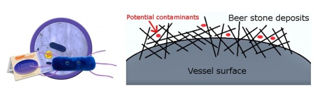
So, while in the body, if the bacterial cell attaches to a surface, it will begin secretion of diphtheria toxin. Interestingly the amount of toxin released in the infected person is dependent on two factors. Firstly, the level of iron outside of the cells in the tissue of the respiratory tract, acts to repress the amount of toxin released. The more iron present, the less toxin released. Secondly, the gene for this toxin is actually found on a virus (beta phage) that can be integrated with the bacterial cell, as opposed to being included as part of the bacterial cell’s machinery. So, once the virus is integrated into the bacterial cell, the toxin can be released.

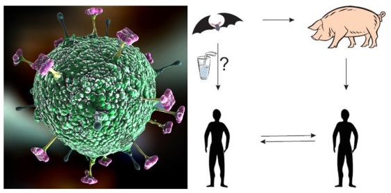
Even though toxin release is dependent on a virus and iron levels, C. diphtheriae has a fast doubling time, and seems to be quite detrimental to humans, especially in the developing world. Interestingly as well, mice seem to be immune from infection, so scientists need to use another model to study this disease. One can find C. diphtheriae in the mouth, skin, nose and throat of infected people, and as a result, this contagious disease can be spread through the air via sneezing or coughing.

As of late last year, cases of diphtheria and several fatalities were reported, and often times children are affected. Vaccination has been a good approach, as well as use of antitoxin and antibiotics. Hopefully in the near future, there can be eradication of diphtheria. Who knew the combined influence of a virus and iron could be so important!

Take care till next time! 🙂

Follow the links to read more on diphtheria, C. diphtheriaediphtheria microbewiki and diphtheria history. Credit is given for use of image of C. diphtheriae and big sneeze.

Tobacco mosaic virus…the most studied of plant viruses!

Hello everyone and welcome back! My cousin’s birthday was Christmas day and sometimes for birthdays and other occasions, people often get flowers or plants as gifts. Well, all her plants are doing well, but sometimes plants can get sick, and could be due to viral infections…just like humans!

Blog 12.31.17

Viruses can be very specific, however, tobacco mosaic virus (or TMV for short), is able to infect a variety of different plants…more than 100 different species of plants to be exact! TMV was the first virus to be discovered, and can cause characteristic blotched spots on leaves and other parts of the plant. The effect of the infection is dependent on the type of plant it infects. This virus is a single stranded, RNA virus that contains subunits coiled in a cylindrical structure. If you are a student, can you quickly research which Russian botanist did significant research on TMV and who is credited as the discoverer of viruses?

The virus itself is very stable (one of the most stable viruses known), and can survive temperatures of 50oC on dry leaf matter. TMV moves from adjacent cell to cell in the plant, with the virus being transmitted to the plant through handling by humans. A good way to prevent infection therefore, is to use proper sanitation practices such as hand washing between handling different types of plants. Another way to avoid infection is through crop rotation of at least a 2-year period.

Since it infects so many species of plants, it is of great importance from an agricultural perspective. Much research has been and continues to be done on this virus, making it the most studied of plant viruses!

Take care until next time and happy new year!! 🙂

Follow the links to read more on Tobacco mosaic virus link1TMV link2, and Dmitri Ivanovsky. Credit is given for picture of infected leaves, TMV in a top hatinfected tomatoes, and the TMV structure.

Shaped like a comma.

Hello everyone and welcome back! The semester has just ended, which means that I’ve finished teaching my class about Vibrio fischeri related information (they were a good group, so I’ll miss teaching them)….however, there’s another species of Vibrio I’d like to talk about this week.

Blog 12.17.17

I remember growing up in Trinidad, and seeing ‘Stop Cholera’ advertisements on television. They would encourage people to boil water to avoid cholera outbreaks. Cholera itself is a disease caused by the gram-negative bacterium named Vibrio cholerae. The organism resembles a comma, with a single flagellum located at one pole. If you are a student, can you quickly research which Italian scientist discovered this organism back in the 1800s?

It was once thought that cholera was caused by something in the air, however, this was found to not be true at all. There are actually different types of Vibrio cholerae…some are pathogenic (causing disease), while some are not. Cholera is identified through acute diarrhea, muscle cramping and vomiting. It only lasts a few days, but those days of sickness can’t be pleasant. English physician John Snow (not to be confused with fictitious character Jon Snow from Game of Thrones!), conducted much research on cholera, leading to advancements in topics on human health and disease.

So, how does V. cholerae work? Well, they get inside us through ingesting contaminated food or water. They use their flagellum to guide and propel them through the mucus of the small intestine. Once they get there, they switch off their mechanism for using the flagella, so that they can channel that energy elsewhere (yes, I know…quite clever really). They then start to produce toxin, which leads to the production of the watery diarrhea.

The last big outbreak was recorded in 2010, and if treated quickly, the mortality rate can be kept quite low. Taking fluids, antibiotics, zinc supplements, and electrolytes are modes of treating affected persons. In Bangladesh, using sari to filter water is an ingenious and inexpensive method they’ve used to decrease the incidence of cholera.

Interestingly, there is a non-toxic subunit of the cholera toxin itself, that is used to detect neurons within the body. If labeled with a fluorescent marker, this can be used during operations that may require this sort of help.

So, general good sanitation (including handwashing), and purifying water for drinking or cooking (either by adding chlorine, filtering or boiling) are ways in which cholera can be avoided from this organism that’s shaped like a comma.

Take care until next time! 🙂

Please follow the links to read more on choleraV. cholerae (link 1), and V. cholerae (link 2). Credit is given for using images of V. cholerae electron micrographV. cholerae giant microbe and sari filtration.


A spring-loaded poison dagger

Hello everyone and welcome back! In last week’s blog, I spoke a little about enterohemorrhagic E. coli…a.k.a. EHEC. This organism possesses components that can affect the way in which it develops the disease it causes (pathogenesis). This week I’m going to talk about one of these components, namely a Type 6 Secretion System (T6SS).

Blog 12.10.17

EHEC is an emerging pathogen that causes bloody diarrhea in patients. It is a foodborne bacterium, acquired by ingesting contaminated foods. Human infection has resulted from direct or indirect contact with contaminated ruminant feces. If you are a student, can you recall what you can do to prevent EHEC infection?

The T6SS itself is used by gram-negative organisms to move/transport proteins from within the bacterial cell, into an adjacent target cell. This system was only fairly recently identified in Vibrio cholerae. The structure itself is said to look like an upside-down phage (a virus that infects bacteria), and extends outwards from the surface of the bacterial cell. It possesses several proteins placed together into 3 sub-complexes. These are a base, a tiny tube (tubule) that resembles that of a phage tube structure, and one other that covers the distance across the cell membrane (a membrane-complex). These complexes collectively move proteins to a close-by target cell, via contraction. The tubule assembles and disassembles several times, can be up to 600nm long, and pictures taken with an electron microscope can capture their structure. The membrane-complex anchors the system to the cell membrane, providing a channel for substrates to pass.

So, I found a good video that provides a visual of the T6SS at work, in V. cholerae. Here, they describe the T6SS as a poison dagger, and narrate some research that was published in Nature in 2012. The researchers labeled one of the proteins with a fluorescent dye so that they could see that the T6SS is a lengthened structure in living cells. It quickly assembles, contracts then disassembles. Assembly is said to occur within tens of seconds, but firing takes less than 5 milliseconds! Cryo electron microscopy (a form of TEM that uses very cold temperatures) was used to see this more closely. The tubular component anchored to the cell membrane is the T6SS itself, and so, this inner tubule is pushed out of the cell, following contraction.

Further research has been done on T6SS since the above-mentioned publication. Some work even suggests that the EHEC T6SS may be able to increase EHEC’s survival, while engulfed within specific cells (macrophages), by counteracting compounds released by these macrophages that aim to kill pathogenic bacteria. Quite clever for a spring-loaded poison dagger don’t you think?

Take care until next time! 🙂

Follow the links for more information on T6SS and pathogenesis. Credit is given for use of Cryo-electron micrograph of the T6SS, and T6SS structure.

A little about EHEC

Hello everyone and welcome back! This past week I helped discuss a scientific paper that focused on a strain of E. coli that causes health concerns in animals…including us! In this blog, I’ll talk a little about one of these strains.

Blog 12.3.17

What do you know about E. coli? Well, they are gram-negative, coliform bacteria that tend to be found in the intestines of warm-blooded organisms. Some strains are harmless, but others are pathogenic (cause disease). The harmless ones could help protect the body from colonization of pathogenic strains. When we say coliform bacteria, we are talking about rod-shaped gram-negative bacteria that could ferment lactose to make acid and gas at temperatures between 35-37oC. E. coli is readily found in fecal matter, and quickly multiplies, within days, in the presence of oxygen.

The major way to spread pathogenic strains is therefore through the fecal-oral route. Lucky for us, E. coli has been studied for decades, is easy to grow and reproduces in about 20 minutes. E. coli itself is one of the most diverse bacterial species, is able to transfer DNA, allowing for horizontal gene transfer. Horizontal gene transfer refers to movement of genetic material from one organism to another, rather than from a parent organism to offspring. Specific processes allowed for the movement of shiga toxin from Shigella to E. coli strain O157:H7. This strain is also called enterohemorrhagic E. coli…or EHEC for short.

EHEC in particular is a foodborne bacterium and so, is acquired by ingesting contaminated foods. EHEC was isolated from hamburger meat, and it has been found that direct or indirect contact with contaminated ruminant feces to be the culprit of human infection.

This bacterial strain is motile, and uses rotating flagella to help navigate through the mucus of the GI tract. The bacterium senses that it’s in the environment it wants to be in (pretty smart right?), and gets ready for attachment by producing fimbriae, which are essentially attachment pili, that are short and thinner than flagella.

So, we have spoken specifically about EHEC, but you have many flavors of pathogenic E. coli to choose from. Some of these act on the intestine, while others act outside of the intestine, causing a variety of diseases. These include good old diarrhea, dysentery (which is diarrhea with blood…may include fever and stomach pain), kidney inflammation, colitis (inflammation of the colon), meningitis (inflammation of the protective membranes protecting the brain and spinal cord), and septicemia/bacteremia (presence of bacteria in the blood). If you are a student, can you find the name one other strain of pathogenic E. coli?

So, why do we care of about EHEC? Well, it is an emerging pathogen, causing bloody diarrhea in patients, and affects the young and old in particular. Any food source can transmit this pathogen (it even adheres well to leaf surfaces), and low infection doses can help explain person-to-person transmission, as well as animals to humans. An infected person can shed up to a billion EHEC cells per gram of feces, even though starting with an infection dose of as little as 10 cells! The size of outbreaks has tended to decrease, with the relative number of outbreaks having increased since the 90s, although the number of infections seems to be stable.

A good way to avoid infection is by practicing good sanitary practices (such as washing hands after using the restroom, and washing hands before preparing food). Additionally, it is good to wash vegetables, and cook meats well.

Take care until next time! 🙂

Please follow the following links for more on EHEC, and horizontal gene transfer. Credit is given for use of images of E. coli giant microbehand washing and washing vegetables.

Microbes in very cold regions

Hello everyone and welcome back! Last Monday our department hosted a seminar speaker, Dr. Lyle Whyte, who studies microbes in very cold regions. One such organism is Planococcus halocryophilus. I hadn’t heard much about this interesting organism, so in case you haven’t as well, I decided to share a little with you this week.

Blog 11.26.17

This microbe, Planococcus halocryophilus originates from soil found from Canadian high Arctic permafrost. Permafrost refers to soil, ground or rock that is pretty much permanently frozen (frozen for at least 2 consecutive years or more). There may or may not be ice present on the ground in these places. This environment is quite extreme when considering existence of living things. Thus, organisms that have adapted to live in these extreme environments are called extremophiles. If you are a student, can you think of any other places on the planet where you can find extremophiles?

Now, cells, for example those found in you and I, undergo a series of chemical activities that are needed to sustain life (a.k.a. metabolism). This includes creating food, allowance for growth and reproduction, as well as removal of cellular waste materials. One incredible thing about P. halocryophilus is that it is able to grow and divide at temperatures as low as -15oC/5oF! For comparison, if our body temperature drops to less than 20oC/68oF, we show signs of severe hypothermia, and no vital signs are present. Normal human body temperature is maintained at 36.5-37.5oC and hypothermic conditions begin when this temperature drops below 35oC. For further comparison, P. halocryophilus can still sustain life at -25oC/-13oF!

Dr. Whyte and his group have thus found an organism that they have shown can live at such extremely cold temperatures. This gram-positive, circular shaped, motile bacterium has indeed set the record for survival at the lowest temperature (-15oC) currently known for growth of bacteria.

For those interested in what life may be like in other parts of our solar system with extreme environments, for example on Mars, this bacterium may just be able to give us some important insight.

Take care until next time! 🙂

Follow the links to read more on research from Dr. Lyle Whyte, hypothermia, metabolism., extremophiles, and permafrost. Credit is given for use of P. halocryophilus imageMars image and image/map of the Canadian high Arctic.

An untapped resource

Hello everyone and welcome back! Well, there was too much to share from the career based panel from last week’s American Society for Microbiology (ASM) conference, so I have a little more to share with you this week.

Blog 11.19.17

Any water that has been negatively impacted by human usage is defined as wastewater. So…how much do you think about your wastewater and how much you pay to have it treated? Well, maybe not a whole lot. One speaker addressed the way in which there is synergy between microbiology and engineering in terms of wastewater. If you are a student, can you quickly check how much money is spent in your household to take care of your wastewater?

Wastewater requires biological treatment. So, the understanding of this type of treatment, and the biological processes that are a part of it, have been needed for chemical engineering for more than 100 years. From an engineering perspective at least, there is an effort to change the way in which we think about wastewater. Actually, there is so much carbon, nitrogen and phosphorus associated with wastewater! For instance, if carbon = energy, one such way to use this carbon is by trying to harness energy from this waste source.

Additionally, phosphorous is a non-renewable resource that is used as fertilizer to grow our food…and we maybe have another 100 years until this resource is depleted. This means therefore that the cost of fertilizer will increase, as well as the cost of food. As a result, there is an effort to try to identify natural microbes that can harness phosphorus.

Unfortunately, there is not much funding allocated for this sort of research, and so innovation has to be done as efficiently as possible. For example, ammonia can be converted to gas without investment of a lot of energy. In terms of energy efficiency, microbes called ammonia oxidizers can produce 4.5 grams of oxygen for every gram of ammonia! One example of an ammonia oxidizing bacterium is Candidatus Scalindua sp., which can be found in marine environments.

ANaerobic AMMonium OXidation, or Anammox for short, describes a nitrogen cycling process mediated by specific bacteria. Here, ammonium and nitrite ions are converted to nitrogen and water. At the moment, there is interest in engineering a process to make Ammonox bacteria happy, to be able to more efficiently produce nitrogen gas, and therefore decrease the cost of your sewage bill. Wouldn’t that be great?

Much work is therefore required from a microbiology and engineering perspective, showing that wastewater itself can therefore really be considered an untapped resource.

Take care until next time! 🙂

Follow these links to learn more about wastewater, and Anommox. Credit is given for use of image of wastewater treatment plant, phosphorus, nitrogen, carbon and Anommox. Candidatus Scalindua sp.

The best of two worlds! Micro style

Hello everyone and welcome back! This past week the American Society for Microbiology (ASM) held a regional meeting in Cookeville Tennessee…not too far away from Knoxville. Many of my peers in the department gave talks and poster presentations, and two topped the prizes for best and third best talk! (Go Vols!). There was an additional career based panel during lunch on the second day, and I will share a little bit about what was said then.

Blog 11.12.17

One panelist spoke about two jobs that he currently holds, that both use microbiology knowledge. One job included responsibilities at a food industry-type setting, while the other at a microbrewery.

In many industries, for example those concerning poultry, microorganisms such as Listeria like to grow and be problematic. This rod-shaped, gram-positive microbe causes the infection called listeriosis, which affects the central nervous system and gastrointestinal tract. Persons who are immunocompromised may also see signs of bacteria in the blood (called bacteremia). Listeriosis occurs when contaminated food is eaten. If you are a student, can you quickly research which surgeon this bacterium was named after?

In general, companies are trying to ‘clean up’ their labels, with respect to what compounds are being used as preservatives, and so there is an interest to have all natural products that are antimicrobials. As a result, bacteria are being grown for their natural antimicrobial products. It was mentioned that some antimicrobials can actually be produced from sawdust. An interesting point made was that pet food is a high-end market that is also trying to get ‘cleaner labels’.

At the microbrewery job, this panelist mentioned that hops itself is greatly used for its antimicrobial use. In this business, it is very important to control temperature and the variety of products that yeast make. Yeast have nutritional needs, and there are a vast number of types of yeast. The ones used for beer are very specific, and it is very important to know how many cells are present. Why, you ask? Well, there are smells and attributes produced while the cells grow, which include aldehydes and phenols. Therefore, cell counts are necessary in controlling these. Interestingly, the number of living to dead cells needs to be known, because the dead cells can release bad compounds (although some dead cells can add nutrients). Another interesting point was that ‘your worst enemy’ is the production of biofilms (called beer stone). This may cause minor taste issues, or major problems. This biofilm can create an environment for microorganisms or contaminants to hide, and it is always better to not have beer stone than to remove it.

Therefore, there is scope for microbiologists in food industry settings. Also, in case you are interested in a (microbiology) career at a microbrewery, there are classes and training that can be done to pursue this passion. If you want, by example of this speaker, you can have the best of two worlds!

Take care until next time! 🙂

Please follow the links to read more on Listeria, bacteremia, listeriosis, beer stone. Credit is given for use of the giant microbe Listeria, and beer stone image.


A virus that inspired Hollywood!

Hello everyone and welcome back! This past week I was reading some scientific literature and I came across a virus I hadn’t heard of (yes…I was surprised as well!). As I learnt a bit more about it, I decided to share a little about Nipah virus with you all.

Blog 11.5.17

Once upon a time in Malaysia, back in 1998 to be more precise, there appeared to be an outbreak of persons suffering from inflammation of the brain (encephalitis), and it was realized that Nipah virus (NiV) was the culprit. This virus has the ability to hop from one host species to another (called a zoonotic disease), and seems to have a reservoir in Pteropus fruit bats in Malaysia. Pigs were found to be an intermediate host, and humans can get infected from infected individuals from either species, or from an infected human. If you are a student, quickly research another example of an infamous zoonotic disease that has been particularly problematic in the past few years.

Why these reservoirs of the virus are greatly worrisome to researchers is that bats can migrate over fairly large geographic locations, while pigs are reared and greatly transported in that region. NiV is apparently becoming of increased concern in Australia and even North America. There is no vaccine for the virus, and it can be detected by analysis of tissue, for example in the kidneys and liver. This virus has a very high mutation rate which makes it difficult for a vaccine to be created in the first place.

In addition to Malaysia, Singapore and Bangladesh have seen the likes of several outbreaks of this virus, with cases in Bangladesh being reported as recently as 2013. Much about the molecular workings of the virus remain unknown, as well as how long NiV could survive outside a host.

So, if by chance you have watched the movie Contagion, fun fact…it was this virus that supplied the inspiration for the virus discussed in that movie. If you have not watched it, maybe you can have a look soon, and think that you now know more about a virus that inspired Hollywood. 😉

Take care until next time! 🙂

Check the following links to read more on Nipah virus and encephalitis. Credit is given for use of images of a Nipah virus model and  virus transfer schematic.

Sometimes more is not better

Hello everyone and welcome back! This past week I was chatting with a former student, and during that time she reminded me of her love for jellyfish. In thinking about the marine environment, I wondered about the relationship between jellyfish and bacteria, and how their lives may be intertwined.
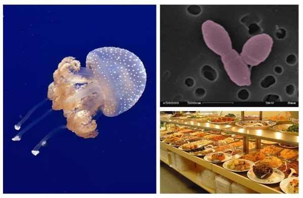
Blog 10.29.17

So, my research focuses on the relationship between specific marine bacteria and the viruses that infect them. The bacteria that I’m interested in are all part of a big group (or Class), called Alphaproteobacteria. When bacterial cells burst open or essentially die, organic carbon is made available to other organisms around them. So…what happens when organic carbon is released into the marine environment from jellyfish?

Well, it seems as though increased funneling of a variety of pollutants and introduction of man-made debris into the marine environment, overfishing, increased water temperatures, and other such factors, create ideal conditions for jellyfish blooms. This is because jellyfish prefer warmer waters, and instances such as overfishing remove competitors for food. We also have to consider that jellyfish have limited predators. If you are a student, can you quickly research two organisms that prey on jellyfish?

So, when jellyfish die, all their mucus and gelatinous material can become nom noms for organisms such as bacteria. Sounds like a great food party for marine bacteria, right? Maybe not so fast!

My close friends and family know how slowly I eat….and that I could only eat so much at a time. If I get 1 chocolate-chip cookie, or even 2, I’d be happy. If you give me twenty however, as much as I’ll be happy and even give you a hug, I can’t eat that much. Worse yet at a buffet…I just can’t eat several plates of food!! Well, something similar happens to the bacteria in the jellyfish scenario. Even with a large amount of carbon rich organic matter available, the marine bacteria are not necessarily growing or reproducing faster, and much of the carbon ends up somewhat shunted/lost from the ocean food web.

What is also interesting is that researchers have found evidence for shifts in bacterial composition when fed organic matter from jellyfish. It seems as though the amount of Alphaproteobacteria (the Class of organisms that I study, and the dominant group in the study), decreases, while another Class called Gammaproteobacteria increases.

Changes to the marine environment may indicate an increased number of jellyfish blooms, and thus changes to community structure and food webs. In general, this highlights the importance of marine bacteria in biogeochemical cycling. However, if jellyfish blooms become more frequent, and in terms of amount of available organic matter, maybe sometimes more is not necessarily better.

Take care until next time! 🙂

Follow the links for more on jellyfish shifts, Tinta et al. 2012Alphaproteobacteria, and Biological classification: Class. Credit is given for use of images of the Australian spotted jellyfish, Jannaschia sp. , and a buffet.