Shrimp white spot syndrome

Hello everyone and welcome back! Muppet treasure island is a movie I like a lot (you should check it out if you haven’t already haha), and in it there’s reference to the ‘black spot’. To a pirate, the black spot was a bad sign, and the recipient of said spot knew that they probably wouldn’t be alive for much longer. Well, today I’m going to talk about another spot, that is just as bad of a sign for some shrimp.

Blog 4.22.18

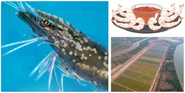
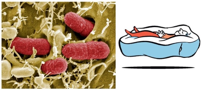
Whispovirus, a.k.a white spot, is the only genus of virus in the family Nimaviridae. This virus contains DNA and actually, a group (or complex) of viruses, called the Whitespot Syndrome Baculovirus complex, causes the disease called white spot syndrome. This virus particularly infects a specific type of shrimp (penaeid shrimp), although it can also infect other types of crustaceans. Characteristic white spots are created on the shell of the shrimp…and they also get sluggish and lose their appetite. We care about penaeid shrimp because many of them are consumed as food around the world, and a lot of money passes through many hands because of this industry. If you are a student, can you quickly research what other type of crustacean this virus affects?

In the early 1990s, there were reports of huge white spot outbreaks in Taiwan and China, causing a big problem for the shrimp farming business. Several other countries such as the United States, Saudi Arabia, India and Japan have all seen instances of outbreaks, with reports being made as recently as 2016 in Australia. What further makes this virus such a problem is that there is no current treatment for the disease, it is very contagious, it kills all the shrimp in a given location, and it does it fast! So fast in fact, that all the shrimp in a given shrimp farm could be wiped out within days!!

Well then…what can we do about this problem you ask? Prevention is key. So, it appears that penaeid shrimp like a relaxed life (like some people I know…haha), so, it’s a good idea to keep reared shrimp away from environmental stressors, which include big changes in temperature or salt content, or exposure to bacterial infections. Disinfectants can be used, and farmers are careful to avoid instances of contamination. Overall, it seems as though much research still needs to be done (hint hint to any budding scientists out there), to help discover treatments for shrimp white spot syndrome.

Take care until next time, and happy Earth Day! 😊

Follow the links to read more on white spot syndrome, and Penaeidae. Credit is given for use of images of white spot diseased individualMahajamba shrimp farm, and shrimp with sauce cartoon.


Wilting away

Hello everyone and welcome back! A couple months ago a friend gave me a plant, that I’ve now named Eugene (don’t question it too much…the plant just looked like a Eugene haha!), and ‘he’ seems to be doing quite well and suffers from no bacterial infections (…that I know of!). However, there are some plant species that are susceptible to bacterial infection by the gram-negative pathogen Ralstonia solanacearum, and I’ll talk a little about this organism this week.

Blog 4.8.18

R. solanacearum can be found not only in the tropics and subtropics, but it is also problematic in temperate areas. This bacterium can cause infection in more than 200 plant species, and loss of plants is estimated to be in the hundreds of millions of dollars every year!! Do you cook with potatoes or tomatoes? Like to plant Geraniums? Or even like bananas? Well, R. solanacearum can infect them all!

This bacterium can be found in the soil, and there just needs to be at least one opening point for R. solanacearum to enter the plant. The bacterium is motile, and can actually sense and move towards the plant itself. A plant wound may be naturally formed as the plant grows, or by animals such as bugs or nematodes, or through every day agricultural activity.

The bacterium colonizes the plant’s internal water system (the xylem), and causes wilting of the leaves. Essentially, as more bacteria start to block parts of the xylem, it disrupts the natural flow of water, and the leaves don’t get all the water they need. If cut, an ooze can be seen leaving the stems, which could lead to bacterial contamination of the soil, and even agricultural equipment. R. solanacearum could also spread through surrounding water, insects, irrigation methods and seeds. If you are a student, can you quickly research which American plant researcher proved that this disease was caused due to bacteria?

So, this particular bacterium is detrimental because it infects so many species of plants in a wide geographical area, is quite lethal, causes great economic loss, and can survive in the environment (under a variety of conditions) for many years. So…what can we do to overcome this problem? Well, it’s always a good idea to use agricultural equipment that are not contaminated. Also, it can help to conduct crop rotation practices, using plants that are resistant to infection, following those that are susceptible. Depending on the extent of the problem, eradication and sanitation efforts may need to take place.

Research has been done on R. solanacearum, but much more is still ongoing to understand the workings of this bacterium that causes this plant wilting away.

Take care until next time! 🙂

Follow the links to read more on R. solanacearum, Geranium, and Erwin Frink Smith. Credit is given for use image of Erwin Frink Smith, wilting in a tomato plant, and brown rot in a potato caused by R. solanacearum.

Not always a great party in the sea!

Hello everyone and welcome back! Many of my friends know that I love the ocean and anything related to the sea. Yes…growing up, I was even one of those girls who owned dolphin pendants and had even gone to see Leatherback turtles lay eggs on one of the beaches in Trinidad. Well, sometimes it’s not always a great party in the sea, and this week I’ll write a little about a dilemma some sea turtles face.

Blog 3.25.18

Sometimes, sea turtles develop tumors, believed to be caused by a type of herpesvirus. The family of herpesviruses (Herpesviridae) contains DNA and some types can cause infection in humans! For example, if you’ve ever had chickenpox, your body now carries just one of a variety of herpesvirus that can infect people. If you are a student, can you quickly research what is the origin of the word Herpesviridae?

Overall, there are more than 100 types of herpesviruses that infect many different animals, and sometimes antiviral agents can be effective. The disease with these tumors, that develops and is specific to sea turtles, is called Fibropapillomatosis (or FP for short). Although FP can be seen around the world, it is much more severe in warmer climate waters (such as the Caribbean and Hawaiian Islands). Young turtles are more prone to FP, and although there are external tumors that are very obvious to the eye, some internal ones may also exist. Depending on the extent of tumor development, these young ones may not live to see adulthood.

Although sea turtles can survive much physical battering, they are much more susceptible to problems started from a human source, such as specific contaminants. The presence of these contaminants/toxins, different stresses, and increasing water temperatures, can be factors that affect and influence FP (and virus) prevalence.

Some evidence links the overall quality of sea turtle habitat as an influence for seeing tumors…the more pristine the waters, the less likely the presence of tumors on the turtles. Maybe there can be little (or big) things that we as humans can do to help these pretty awesome marine creatures.

Take care till next time! 🙂

Follow the links to read more on sea turtle fibropapillomatosis, Herpesviridae and Varicella zoster virus. Credit is given for use of image of sea turtle with tumors sea turtle with tumors, Trinidad and Tobago turtle habitat, dabbing sea turtle.

Lactobacillus plantarum in the spotlight

Hello everyone and welcome back! I’ve recently had a craving for popcorn (cannot explain that one…), but it had me think about gut bacteria on a whole. Well, one bacterium species, Lactobacillus plantarum was mentioned during a recent seminar, and how it helps the gut. I’ll talk a little about this bacterium this week.

Blog 3.11.2018

Lactobacillus plantarum is a gram-positive bacterium that is naturally found in many foods (does anyone reading like brined olives? How about pickles? L. plantarum could be found in these 😊). L. plantarum was first found/isolated in saliva, and can survive in a wide temperature and pH range. This species can also dissolve gelatin (fun fact). If you are a student, can you quickly research one other species of Lactobacillus that may be important to us?

So, interestingly, this bacterium species is able to respire oxygen…but they don’t have the ‘equipment’, so to speak, to carry out that process. Usually, in cellular respiration, carbon dioxide is a resulting by-product. Here, hydrogen peroxide ends up being a final product. What this does, however, is stop the growth of other competing microbes that cannot survive in the presence of hydrogen peroxide.

Since L. plantarum is often found in many foods, particularly of the fermented variety, it makes itself a promising choice with respect to probiotic development and research. In terms of therapeutic ability, L. plantarum can control the growth of microbes that produce gas in the gut, and can therefore be useful to patients who suffer from irritable bowel syndrome.

I think the mere uniqueness of L. plantarum (…and that it encourages a happy tummy environment haha), makes it worth being in the spotlight this week. 😊

Take care until next time! 😊

Follow the links for more reading on L. plantarum, and cellular respiration. Credit is given for use of probiotics cartoon, happy tummy image, Lactobacillus plantarum image.

A bug with a ‘super power’

Hello everyone and welcome back! Just as there are superheroes in the Marvel Universe, for example, it may seem as though there are bacteria in our universe with super powers of their own. Today I’ll talk a little about Geobacter metallireducens, and what makes it super 😉.

Blog 2.25.18

This gram-negative bacterium is able to move (motile), and only likes to live in places that have no oxygen (anaerobic). Geobacter metallireducens can also move and respond to the availability of nutrients…to places where nutrients are less favorable, to places where they are more so (called chemotaxis). Through specific chemical reactions, G. metallireducens can alter fatty acids and other compounds by using iron. If you are a student, can you quickly research what these important chemical reactions are called?

So, what’s so super about them? Well, this organism, first found in freshwater soil, are able to use uranium, and plutonium, to grow. Not only this, it can draw power/energy from uranium and other metals. It can also take up/use harmful compounds (which may even be radioactive), and make them harmless. Its ability is so important in helping clean up geographical locations that are contaminated with these sorts of compounds. You wouldn’t want uranium in any of the water you drink, now would you?

Studies have been done to look at what genes are included in this organism, and how it functions. There is also application, in the realm of bioengineering and biotechnology application, for persons to study this bug with a ‘super power’.

Take care until next time! 😊

p.s. this post makes my two-year anniversary of science blogging (woohoo!). Thanks so much to all those who have read my blogs at some point during this time. It means the world to me 😊.

For more on G. metallireducens, and some more bugs with a ‘super power’, see this video. Also, read more on Geobacter metallireducens, chemotaxis, and Redox reactions. Credit is given for use of ‘super bacteria’ image and Geobacter sulfurreducens giant microbe image.


One of the big guys!

Hello everyone and welcome back. Well, as you could imagine, bacteria come in all different shapes and sizes. Some of them such as E. coli strains be around 1-2um in length. The bacterium I’m going to talk about this week makes E. coli look quite tiny.

Blog 2.18.18

Epulopiscium fishelsoni…or Epulo for short, can be 10-20 times longer, and can have a volume of more than 2,000 times that of any other average bacterium. It is because it is so unusual, it was once put in the protist kingdom for classification, rather than bacteria.

It appears that not only its size is unusual in compared to other bacteria, but also its reproductive cycle. Several daughter cells could be held within the parental cell, until the already mobile daughter cells burst out into the environment.

Interestingly, Epulo was isolated from the gut of brown surgeonfish from the red sea back in 1985. If you are a student, can you quickly check which other places since then, Epulo has been isolated?

Epulo has been shown to alter the metabolism of its host…daily! While the fish is active and feeding during the day, the Epulo cells lengthen, while suppressing the pH of the fish gut fluids. During the night, the Epulo cells get shorter and there is no pH suppression.

While there are other oddities of Epulo, the differences of its daily life cycle introduce difficulties (near impossibilities really) in having them grow in lab culture and conditions.

This unicellular bacterium is definitely one of the big guys!

Take car until next time! 🙂

Follow the link for more on Epulopiscium fishelsoni. Credit is given for use of Epulo image.


Pretty cool

Hello everyone and welcome back! In the US there have been waves of cold weather, creating snow storms, at some locations, in the process. Well, though some like it cold, this Caribbean girl would prefer some warmer weather soon haha! So, as much as it’s fun looking at snow on a macro scale…what if I told you that there are microbes that may help create it? This week, I will therefore speak a little about Pseudomonas syringae.

Blog 2.11.18

The gram-negative bacterium Pseudomonas syringae is rod-shaped, can move, and infects plants. There are several strains/types of this microbe (around 50 in fact…with each strain affecting each plant species differently), and gets its name from the tree species from which it was first found (Syringa vulgaris…a.k.a. the lilac tree). If you are a student, can you quickly research who isolated P. syringae back in 1902?

Since some strains are plant pathogens (causing disease in plants), these can secrete a toxin called syringomycin, that affect plant cell membranes. P. syringae can be found colonizing the surface of leaves (known as the phyllosphere), but if pathogenic, they end up destroying their ‘homes’, by forming lesions, for example.

But here’s where it gets very interesting…this microbe is able to create its own ice! Yes, you read that correctly haha. They are living ice makers. They have surface proteins that imitate ice crystal structure, encouraging attachment of more water molecules to them. When this happens, frost damage is possible on plant tissue when temperatures are a little below freezing. There is a quite amazing video that shows water freezing almost instantly when these microbes are added it!

What’s more interesting, is that P. syringae seems to have a greater influence on snow formation, and other forms of precipitation, than previously thought. Additionally, this microbe is used to even create artificial snow at some snow resorts! Pretty cool, don’t you think? (no pun intended 😉)

Take care until next time! 😊

For more information on P. syringae, see link 1, link 2 and link 3. Also see the following link for more on syringomycin. Credit is given for use of images of icy plant leaves, ski location and P. syringae

Sea foam

Hello everyone and welcome back! I was recently asked about something that was seen along a seashore, and I immediately knew it was sea foam. What I didn’t know was why and how it forms, and I’ll share a little about that with you this week.

Blog 1.28.18

It appears that sea foam is the result of a significant amount of dissolved organic matter that gets churned in the sea water. This type of dissolved organic matter may include complex plant derived compounds called lignin, and/or proteins or lipids. Just as dish washing soap can produce bubbles, these compounds can do the same, creating bubbles in the crashing waves. Often times, these compounds can come from algal blooms that occur offshore. If you are a student, can you quickly think of examples of algae?

Since this sea foam can stick around for a long time, strong sea winds can blow it onto a beach itself. Ordinarily, sea foam is not hazardous to humans. However, if the foam is due to breakdown of harmful algal blooms, direct contact with it could lead to skin irritations. In the case of the motile microorganism Karenia brevis, which causes algal blooms known as red tides, toxins can be dispersed in the air when sea foam bubbles pop. These toxins (collectively termed brevetoxins), can cause respiratory problems in humans (be careful to those who are asthmatic!!) and other organisms (such as birds), that come in contact with it. Additionally, if the source of the dissolved organic matter comes from a polluted water source, or even contains spilt crude oil, the resulting sea foam may be a bit smelly, or may have a different texture.

Overall, it seems that the presence of sea foam often means that the marine ecosystem is a productive one. And now…we all know a little more about sea foam. 🙂

Take care until next time! 🙂

Check the following links to read more on sea foam link 1, sea foam link 2, Karenia brevis. Credit is given for use of  red tide image, Karenia brevis image, and sea foam image.


Of plants and microbes

Hello everyone, and welcome back! I was just yesterday, chatting with a friend of mine about a paper I’m reading for a class. It concerned the relationship between plants and microbes…and then I dropped a word into the conversation…rhizosphere. What is that exactly? Where are the microbes? And what other factors affect plants?? I answered all these questions, and I also choose to explain them here this week.

blog 1.21.18.jpg

Of course, when you look at a plant, there is the part you see above ground…but there is also a whole other world underground! The rhizosphere itself is a very small and specific area of soil surrounding the roots of plants. It is here that the plant roots release specific compounds to the soil, such as sugars, chemicals and other organic compounds. It is in the rhizosphere that you can find many microbes. The microbes can take up nutrients released from the plants, but then microbes can also help the plants by influencing the way they grow, for example. In some cases, such as with legumes, microbes can make nitrogen into a form that the plants can use. Plants and microbes can therefore form a symbiotic relationship. If you are a student, can you think of one type of microbe that lives in a symbiotic relationship with a type of plant?

There can also be plant-plant relationships, in addition to plant-microbe ones. Some plants are quite competitive, and so some of them release chemicals in the rhizosphere (or beyond), that can control the growth of other plants around them.

In addition to these many interactions happening in the rhizosphere (seems like a big party is happening over there!), resources that are essentially of the environment, such as temperature, water and sunlight, affect plants. These kinds of resources are called abiotic factors. Abiotic meaning not of, or from, a living thing. Biotic factors, on the other hand, include bacteria, plants, fungi and other living organisms, that are living components of ecosystems, and affect other parts of ecosystems, communities or even individuals of specific species.

Of course, there is so much ongoing research concerning these sorts of interactions, but much more needs to be known when it comes to the relationships of plants and microbes.

Take care until next time! 😊

Follow the links to read more on the rhizosphere, and abiotic vs biotic factors. Credit is given for use of images of plant root and rhizosphere, plants and roots in soil, and nitrogen fixing bacteria.


The burger bug’s secretion system

Hello everyone and welcome back! Some time ago I wrote about a system called a Type 6 Secretion System (T6SS), that was also described as a spring-loaded poison dagger. If you recall, the T6SS helped move proteins from inside the bacterial cell into adjacent cells. Well, there is another system called a Type 3 Secretion System (T3SS), that is also quite important for bacterial colonization.

Blog 1.14.18

So, I found a video that does a good job at showing how, particularly Enterohemorrhagic E. coli (EHEC for short) colonizes the gut, and how the T3SS helps. Remember, E. coli species that cause disease will enter your body through ingestion (a fecal-oral route)…well, mostly so anyway. EHEC particularly being a foodborne bacterium, can be acquired by eating contaminated foods.

EHEC itself was isolated from hamburger meat (hence the name of the video, ‘getting to the bottom of the burger bug’), and it has been found that contact with contaminated ruminant feces (either directly or indirectly) to be the culprit of human infection! If you are a student, can you quickly research how many kinds/strains of E. coli there are? Hint…the ballpark answer runs the hundreds!

EHEC can move (using rotating appendages called flagella), and this specific property helps the bacteria navigate through mucus of the gastrointestinal tract…a.k.a. the gut. It is aware that it’s in a favorable/favourable location, and gets ready to attach itself there.

There are several genes and pieces of DNA that control the capability of the organism to bring about disease (a.k.a. bacterial pathogenicity). Timing is very important, and bacterial pathogenicity genes need to be turned on at the right place and right time. Some of these genes code for structural proteins of the T3SS itself.

The T3SS starts to assemble at the inner membrane of the bacterial cell, creating the base structure of the secretion system. Once this is done, EHEC can use other external structures to attach to gut epithelial cell microvilli. After contact is made, further gene expression events happen within the bacterium, and export of proteins from the bacterial inner membrane begins.

If you look at the video, you will observe that the microvilli are effaced/erased/disappears from that spot! The bacterial cell becomes partially embodied in the host membrane itself…as though it’s laying down nicely on a foam mattress! This attachment and effacing lesion that is formed in the gut encourages EHEC persistence here. If you have several bacterial cells all doing this same thing, it can interfere with the natural function of the gut cells, thus promoting diarrhea. EHEC releases shiga toxin, which causes bloody diarrhea, and in some cases, kidney damage.

So, in a nutshell, what is the T3SS? It is a protein appendage/attachment, that many gram-negative bacteria have. It essentially is a sensory probe that helps the bacteria detect eukaryotic cells and secrete proteins that help with bacterial infection. There is direct secretion of proteins from the bacterium into the host cell. These proteins help allow the pathogenic organism to survive and hide from the body’s immune response.

Take care until next time! 🙂

Follow the links to read more on EHEC link 1, EHEC link 2 and serotypes. See link to burger bug video here as well. Credit is given for use of image of mattress cartoon and EHEC.