The one named Onesimus

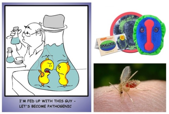
Hello everyone and welcome back! Most people, if not everyone around the world has heard about the disease called smallpox. Luckily for us, the last natural reported case of Variola minor was noted in 1977…now 40 years ago! If you live in North America, specifically the United States, did you know that an African slave’s communication to his master helped save lives from a serious smallpox outbreak in 1721?

Blog 10.22.17

If you have an extra second to google Cotton Mather, you’ll find a webpage, which documents his achievements and status, and even his involvement in the Salem witch trials. Much less is known about his North African slave Onesimus. Actually, where and when he was born…or died…are not known, but he did walk the earth in the 1700s. He was given this biblical name By Cotton Mather who saw promise in him. He lived in Massachusetts at a time where there were not too many people who looked like him, and was able to buy his freedom in 1716, given specific conditions.

Now, in 1721, there a serious smallpox outbreak in Boston. Five years prior, Onesimus had shared knowledge with Mather on an inoculation method…a form of immunization. Powdered scabs or fluid from a pox blister was rubbed into the skin of the uninfected person, who then acquired smallpox naturally with symptoms that cleared up within a month. Today this is called variolation, although vaccines have replaced it.

Variolation is so named because smallpox is caused by one of two types of viruses. These are Variola minor and Variola major. The virus is transmitted through inhalation, invading the respiratory system, and then moving to the lymph nodes. The virus could be found in the bloodstream within two weeks! If you are a student, can you think of another virus that has been of global concern to humans?

Initial symptoms may resemble that of a common cold, however, persons infected with smallpox develop pustules/blisters filled with fluid on the skin. They may even develop limb deformities and blindness. This infectious disease is said to have originated from another animal, and was then naturally passed on to humans, thousands of years ago.

Although Mather believed in, and used variolation, giving Onesimus credit for the knowledge, though many persons who looked like Mather rejected it, because of their apprehensions to African medical approaches at the time. Only two doctors, including Mather used variolation, seeing a much smaller mortality rate than those who were not immunized.

There was no need for the outbreak of 1721 to be as deadly as it was, however more people later saw that this was a good method, and did use it in subsequent outbreaks. As difficult as it was to implement in North America, and gain widespread recognition, variolation had been long used not only in Africa, but in China as well. So here, we give credit to Onesimus for his great contribution, although at the time, he may not have known its impact.

Take care until next time! 🙂

Follow the links to read more on Onesimus, Cotton Mather, Variolation and smallpox. Credit is given for use of giant microbe smallpox and smallpox image.


p.s. special thanks is given to Dr. Jill Mikucki for inspiring this week’s blog and sharing information on this topic. Thank you thank you!


TB or not TB…that is the question

Hello everyone and welcome back! This past week I had done some reading on TB, otherwise known as tuberculosis, a disease most people would have heard of at some point in their life.

Blog 10.15.17

Mycobacterium tuberculosis is just one of nearly 200 species of microorganisms that belong to this genus/group. Mycobacteria need oxygen, are not motile, reproduce very slowly, do not mutate too often, but have some how successfully managed to be around for thousands of years, sometimes causing serious problems in terms of human health and disease. Mycobacterium leprae, is another species from the same genus, for example, and is the organism responsible for causing leprosy in humans. If you are a student, can you quickly research two other Mycobacterium species that may be important with respect to human health and disease?

In talking about slow growth of Mycobacteria…and I mean slow…growth, our good old friend E. coli can go through one division cycle in roughly 20 minutes, whereas M. tuberculosis divides once in roughly 20 hours. M. leprae will take about 20 days! That’s an amazing comparison!

TB itself is just one strain of Mycobacteria, but is actually part of a multipart group, or complex, of different Mycobacterium species. This rod-shaped cell called M. tuberculosis has a cell wall very rich in lipids, and the characteristic acid-fast staining technique is used to observe them under the microscope (as opposed to being described as gram-positive or gram-negative).

What is their strategy do you ask? Well, they infect the lungs, where they are swallowed by special cells called macrophages. Typically, cells that undergo this ‘swallowing’ process are broken down/ killed inside of the macrophages. For TB, this does not happen, and the TB cells multiply. This can happen over several months, or years, and is protected in an inflamed tissue called a granuloma. It is estimated that maybe 1 in 3 people host TB in their lungs, undetected, and the success of the organism is really in how long they can survive this stage in their hosts. When this granuloma breaks down, transmission of TB to other people can occur through infected air droplets.

Although some Mycobacterium species are detrimental to human health, many others are found in the environment (such as in soil), and are much less so.

With so many Mycobacterium species found around the world, together with the prevalence of tuberculosis, it really is a good question as to who may or may not have TB.

Take care until next time! 🙂


For more reading seeing the following links on Mycobacterium,  and Mycobacterium tuberculosis and granuloma. Credit is given for use of lungs image and TB giant microbe.


Methods of microbial control

Hello everyone and welcome back! This past week I stepped in and taught a lecture class (that’s a lot larger than my usual class…mind you…), about microbial control. Of course, microbes can be found in us and on our skin, but also on surfaces in and around the home. Therefore, everyone should know a little about the control of microbes.

Blog 10.8.17

But why? Well, no one likes being sick…nor likes to see microbes growing on our food (when last did you look at a piece of moldy bread and think ‘yummm!’ haha). Based on these two reasons, it is important to understand a little about avoiding food contamination and human illness. We pasteurize milk to avoid bacterial contamination of dairy products, and even use isopropyl alcohol to clean a wound. If you are a student, can you think of two other things that you may use to control microbial growth at home?

In and around the lab we always use aseptic technique, meaning that we make sure to certain surfaces in the lab environment is made and kept sterile. We often use alcohols to keep the lab bench clean, and use a flame to keep some metal equipment sterile. A simple practice of handwashing also tries to ensure that our hands are kept clean.

Whether using a physical or chemical bacterial control in a given location, the way in which they work can be separated into two big ways. They act by either disrupting the outer surface, or the inner components. Think of your home…it protects you from ‘the elements’, and you can get in and out from your front door or side door for example. If someone breaks down a wall, your home is not as protected nor very safe. Alternately, if you have internal components such as central air, or plumbing, that do not work, this can also lead to the malfunction of your home.

Now think of the bacterial cell as a home, and the cell wall or membrane as the walls of the home. Unlike brick or wood, the bacterial cell wall/membrane is formed in a specific way, that allow the movement of substances in and out of the cell. Like a home’s internal components, proteins and nucleic acids are important for other functions of the cell. If the integrity of the cell wall is compromised, the cell bursts open/contents leak out, and if the internal components don’t work well, the cell shuts down/metabolism cannot be controlled.

Of course, in controlling microbes, an ideal control will be cheap, fast-acting, and of no harm to humans, other animals or things…however, an ideal control is hard to come by. Every type of control has limitations, and is dependent on the microbe being controlled, the location of control, and the environmental conditions. Heat, refrigeration, filtration, as well as use of synthetic drugs are all methods of microbial control.

Take care until next time! 🙂

Credit is given for use of  house cartoon.

It’s approaching flu season!

Hello everyone and welcome back. It’s starting to get colder in Knoxville…which means it’s approaching flu season! Therefore, it is only fitting that I mention a little about a paper we recently discussed in a class about the Influenza A virus.

Blog 10.1.17

This paper was called “Emergence of Influenza Viruses and crossing the species barrier”, and was written and published by Koçer and others in 2014. These authors wanted review the rise of viruses that seemingly began and continue to persist in avian species (i.e. birds). They wrote about the crossing of different influenza virus strains from birds to a variety of terrestrial and marine species. There are around 17 known subtypes of flu, where 16 of them are found among birds. Each subtype has a different surface protein called hemagglutin (HA), and it is the H1, H2 and H3 subtypes that have cause pandemic flu in humans. If you are a student, can you remember the name of a strain of flu that was either from birds or pigs that was really problematic to humans in recent history?

So, what is the origin of these flu strains do you ask? Well, it turns out that’s really difficult to say. Since coinfection (infection with more than one strain) occurs fairly frequently, you may have two viruses combining, for example. A new combination of surface toxin (antigens) results, which causes the host organism to respond to this scenario, which would be sickness. This sort of change is called antigenic shift, and is what makes it difficult to determine the origin of these flu strains.

As one would imagine, it would be difficult to have flu strains cross barriers from one species to another. Therefore, it was interesting to note that the number of influenza strains to move from birds to other animals is actually a small amount. Spatial separation, differences in the way in which each species’ body functions (physiologies), and different molecular features (such as the presence or absence of cell receptors for the specific virus), make it difficult to cross species barriers. However, high mutation rates of these viruses greatly enable the transmission. The smallest of changes can result in a more harmful strain of virus.

So, how could there have been a jump of influenza virus from birds to humans? Well, the authors introduce the concept of a mixed vessel. Here, in terms of the evolution of influenza viruses, they mention that swine may play an important role in breaking down the different levels of separation that stop the transmission of these viruses between species. Also, it was interesting to note that swine influenza strains may have even circulated for decades in North America before it surfaces in humans.

Overall, these and other points made in the paper point to the importance of specialists in different disciplines, to carry out continued surveillance (in humans and non-human species), to avoid future country-wide or global outbreaks, and increase disease preparedness.

Take care until next time 🙂

For more reading on Koçer and others in 2014 see Kocer et al. 2014. Also, follow the links to read more on physiology and antigen. Credit is given for use of H5N1 model,  bird flu cartoon and giant microbe bird flu.


The gentle hunter

Hello everyone and welcome back! This morning I had bread and cheese sandwich (nom nom nom)…which, thinking about it, both components involve microbiology to make. So, to address dairy products (specifically fresh milk), and in keeping with writing about female microbiologists, this week’s blog features Alice Catherine Evans.

Blog 9.24.17

Alice Catherine Evans was born in Neath, Pennsylvania, and was home-schooled when she was 5 and 6 years old, and really excelled in her work. She was a basketballer while at the Susquehanna Collegiate Institute, and went on to teach for some years. After teaching, she attained a Bachelor’s degree in Bacteriology from Cornell University by attending free classed offered to rural teachers at the university. She later achieved a Master’s degree from the University of Wisconsin-Madison and was the first woman to get a scholarship for Bacteriology from the school.

She later became the first female scientist to hold a permanent job at the United States Department of Agriculture (USDA), where she studied causes of bacterial contamination of milk products. She also looked at ways to improve tastes of butter and cheese. It is through her interest in the bacterial aspect of fresh, untreated (unpasteurized) milk that she focused on the gram-negative, non-motile, rod-shaped bacterium, Bacillus abortus.

Since this bacterium causes animal miscarriages (for examples in cows), it was hypothesized that human health will be similarly threatened. She discovered that unpasteurized milk from a cow infected with B. abortus, will give rise to Brucellosis (also called undulant fever), in humans. She advocated for the pasteurization of milk. She reported her work and got it published in 1917 and 1918 respectively. If you are a student, can you think about other food products you may get from cows, or even goats?

It is said that her findings were doubted in part because she was a woman and did not have a PhD. In the 1920s however, other researchers from around the world started to come to the same conclusion, and the pasteurization process was later adopted in the United States, greatly decreasing the incidence of undulant fever. The fever has this name because it can come in waves, and can last from several days to many years. Tests on body fluids or blood can confirm the presence of this organism, and treatment is possible by use of antibiotics such as rifampin, gentamicin and tetracyclines. Sometimes a combination of antibiotics may be required.

Unfortunately, after moving on from the USDA, Alice worked at the United States Public Health Service, where in 1922, she became infected with undulant fever (which had no cure back then), greatly affecting her health for two decades. Alice Catherine Evans died at the age of 94, but even after officially retiring in 1945, she continued to encourage women to pursue scientific careers. She received two honorary doctoral degrees, was the first female president of the society of American Bacteriologists, and is an honorary member of the American Society of Microbiology and the Inter-American Committee on Brucellosis.

So, before the gentle hunter went home, she made a great contribution to bacteriology to which we can all graciously say thank you.

Take care until next time! 🙂

Follow the links to read more on Alice Evans, or Brucellosis. Credit is given for use of images of cows, Brucella species, and Alice Catherine Evans.

Microbiology meets forensics

Hello everyone and welcome back! For many months now I’ve been joking with a friend, also pursuing her PhD, but in chemistry, that she can transfer to the microbiology department to do research that interests her. Different disciplines…so big joke, right? Well, it might not be so farfetched.

Blog 9.17.17

Research in chemistry could be quite varied, and my friend’s topic of research is in forensic science, where she’s interested in using superglue (cyanoacrylate) that would stick to fingerprints, allowing them to be seen better. Forensic science refers to the use of science for civil law and criminal investigation. Evidence is gathered, well-maintained and examined throughout the inquiry, and these scientists may be called as experts during these kinds of investigations.

New research is continuing to show that the human microbiome (microbes found on and inside of all of us) can be, and is beginning to be, used in forensic science. Firstly, every person’s microbiome is quite unique, and scientists have been able to even tell identical twins apart based on their microbiome. If you are a student, can you quickly research one type of microorganism that is often associated with your body?

In forensic science, the post-mortem interval (PMI), a.k.a. ‘time after death’, is sometimes difficult to determine. Studies have shown that microbes are everywhere, and will carry out specific processes in a given environment, at particular times. Data, for example from skin microorganisms (which is relatively stable over time), can therefore be used to connect persons to specific locations and times.

Movements forward in microbial ecology research constantly chance the way in which microbes, and communities of microbes, are characterized and understood. One method is metabolomics, which is the study of unique chemical fingerprints that result from processes occurring in a cell.

There is a growing hope that data from individual’s microbiome can tell scientists about what you eat, where you were, who you live with, estimating time after death, and even help in solving crimes! This is a very new realm of study, and microbial data may yet to be accepted for certain forensic evidence. Maybe there’ll be a growing niche where microbiology meets forensics.

Take care until next time! 🙂

Follow the links to read more on forensic science, metabolomics, use of superglue in forensic fingerprinting, Metcalf et al. 2017 review, Microbiome tools for forensic science, ASM article on using skin microbiomes for forensic human identification. Credit is given for images of P. acnes, forensic fingerprints, superglue printing, human microbiome.


The gift of the kissing bug

Hello everyone and welcome back! As promised from last week’s blog, and keeping in the vein of microbial pathogenesis, I wanted to write a little about Chagas disease, and what exactly it is.

Blog 9.10.17

Most likely, if you reside in Latin America, you may have heard about this disease before. A protist by the name of Trypanosoma cruzi is the culprit. By definition, a protist is any eukaryotic organism, which is not a fungus, plant, or animal. T. cruzi is transported to vertebrates, such as humans, through a vector called the kissing bug (or Triatominae). Other members of this subfamily carry names such as vampire bugs and assassin bugs. This subfamily contains more than 130 species, with most feeding on blood from vertebrates (some feed on blood of invertebrates), and tend to be found in close proximity to their blood meals, and predominantly throughout the Americas. If you are a student, can you quickly research which Brazilian scientist this disease is named after?

How do you get this disease you ask? Well, this kissing bug comes out at night, tending to bite sleeping persons on their faces in order to get a blood meal. While doing so, they ‘take a crap’ on the person! (How rude!! And quite gross actually…). The T. cruzi is actually in the feces/faeces itself, left close to the bite spot. When the person starts to scratch the bite, this leads to broken skin and T. cruzi infection. This is very different from how Malaria is spread, for example, where the parasite is introduced to the blood stream, by the mosquito’s saliva, with the bite itself.

Infection with T. cruzi can also occur, however, accidentally in the lab, through blood transfusions, breast milk, or from woman to baby during pregnancy.

This disease can have acute and chronic stages, with no vaccine currently available. Acute symptoms include chagoma (a swelling where the T. cruzi entered the body), fever, nausea and body aches. Immunocompromised persons may suffer greater effects. A notable mark of this disease is called Romaña’s sign, where there’s swelling of the eyelids and face close to the bug bite, and where the feces/faeces was accidentally rubbed into the eye, or was deposited by the bug. Chronic effects include complications of the heart, digestive- and nervous systems.

The disease can be prevented by controlling the vector (the kissing bug) itself by use of insecticides. Medication such as benznidazole can also be used, and tend to work best if administered early.

This tropical disease is the gift of the kissing bug that everyone could definitely do without getting!

Take care until next time! 🙂

Follow these links to read more on Chagas disease, Protists, Triatominae, Chagoma, and Carlos Chagas disease. Credit is given for use of images to show Romaña’s sign, Triatomina, Carlos Chagas, and T. cruzi Giant Microbe.


Origins

Hello everyone and welcome back! This semester I’m enrolled in a journal club class that will meet weekly to talk about scientific papers that pertain to microbial pathogenesis…or in other words, the way in which disease develops. This past week we discussed a paper that addressed the origins of some major human infectious diseases.

Blog 9.3.17

Every piece of scientific literature tries to communicate something, or rather, has a purpose. The purpose of the paper we discussed by Wolfe et al. 2007 (for reference in case anyone reading would like to view it later), was to show that there were differences in the origins of diseases that are/were prevalent in temperate climates, in compared to tropical ones. If you are a student, can you name two diseases that are prevalent in your home town or country?

They suggest that New World (the Americas) diseases came about through the rise of agriculture, and highlight five intermediate stages of pathogen infection of other animals. Remember that a pathogen is defined as a bacterium, or some other microorganism, that can bring about disease. Research is often interconnected with other disciplines, and so this work is not only important for microbiologists, but for historians and evolutionary biologists alike.

Interestingly the persistence of these diseases is dependent on several factors, including rate of infection of new hosts and host population density. The authors specifically looked at data pertaining to 25 diseases, but selected 17 of them based on factors such as current or historical world burdens. Such diseases include HIV, tuberculosis, and smallpox.

Well…what do they find? Here, the differences they see in temperate versus tropical diseases, include vector transmission rates, and type of infection (chronic versus acute for example). The word ‘vector’ refers to another organism that carries a pathogen that can later infect you! Good examples include mosquitoes, or even sand flies. A chronic disease describes one that lasts for a long time. An acute disease is much more short term.

Also, most temperature diseases are described as ‘crowd epidemic diseases’. They see that more temperature pathogens have origins from domestic animals, in compared to tropical pathogens, and may be due to difference in lifestyle (for example in terms of livestock domestication).

Lastly, they seem to address that most diseases that affect humans, are derived from pathogens of other vertebrates, with the exception of two cases of the avian (bird) variety.

In summation, apart from Chagas disease (which I will talk about next week), the diseases of the analyzed database seem to originate from the Old World (Africa, Europe and Asia), rather than the New World (the Americas). This kind of research can ultimately help in the understanding of emerging infectious diseases, as well as its control and detection.

Take care until next time! 🙂

Follow the links to read more on pathogenesis, pathogens, chronic disease, Old World and vector. Credit given for use of sand fly image, smallpox giant microbe and pathogen cartoon.


Curious about caterpillars

Hello everyone and welcome back! This past weekend I went with some friends to a fruit and berry patch. Unfortunately we were too late in the year to pick any berries, and it was too early to pick peaches, so we bought some slushies and decided to take in nature by walking around the farm instead. Then we saw a fuzzy caterpillar going about its business. We didn’t interfere with the caterpillar along its journey, but I then thought, well…what sort of gut microbiome does a caterpillar have?

Blog 8.27.17

Seems as though researchers at the University of Colorado Boulder had the same question. Well, interestingly enough…apparently caterpillars don’t have much of one! The growing idea that all organisms have a gut microbiome doesn’t quite hold true for these guys…and a couple other organisms as well. When analysis of fecal/faecal content were done from caterpillars, then compared to other organisms, caterpillars had roughly 50,000 times less microbes present. This is huge! This stark difference has been described as comparing deserts to rainforests.

The microbes, more so DNA, they do find in caterpillars seems to be associated with that of the leaves they ingest, and their own. Caterpillars don’t seem to need the microbes they have, or rather, there is no consequence in development with or without them. Unlike termites that need to exist in symbiosis with Trichonympha to digest plant material, caterpillars don’t need such symbionts. Symbiosis describes the close and biological relationship between two different organisms. This relationship can be necessary or optional. It can also be beneficial to both, beneficial to one, or harmful to one of the organisms involved. If you are a student, can you research one other example of symbiosis?

It truly is interesting that with the 180,000 caterpillar species on the planet, if they don’t have a gut microbiome…well, what mechanism do they use for food digestion? This is quite new research, and Tobin Hammer, Melissa Whitaker and Jon Sanders are just a few researchers who are on the chase to find out more about this curious case about caterpillars.

Take care until next time! 🙂

See the following links for more on the caterpillar microbiome articleNature news on caterpillar microbiome, symbiosis, and Trichonympha.


Just one slice of cake!

Hello everyone and welcome back! Today’s my dad’s birthday, and I wish I were home to celebrate with him, and maybe get him a birthday cake. Of course that got me thinking about microbes found in the mouth that may be associated with the break-down of carbohydrates found in cake.

Blog 8.20.17

Streptococcus mutans is a round-shaped, gram-positive bacterium (remember…it’s the type of bacterium with the thicker cell wall), that virtually everyone has in their mouth! There are several different species of Streptococci that could also be found as part of normal oral flora, however, this one is a major culprit for oral disease. If you are a student, can you quickly research who is credited for first describing S. mutans?

What this bacterium can do is break-down (i.e. metabolize) carbohydrates such as sucrose, into (lactic) acid. The greater the acidic environment in the mouth, the greater the possibility of erosion of tooth enamel. Have you ever seen plaque on your teeth? Well, when these bacteria grow and multiply, they attach to the surface of teeth (via proteins found on the surface of the cell), and form what is known as a biofilm…i.e. dental plaque. The quantity of S. mutans found in the mouth at a given time has actually been described to affect that of other oral bacteria. Additional research has shown that the type of carbohydrate consumed, along with the age of the host affects specific gene expression of S. mutans. For example, findings helped conclude that younger host animals are more susceptible to the formation of dental caries. Further studies found that much more bacterial activity occurs when host animals had intakes of sucrose, in compared to fructose or glucose.

Of course good dental hygiene, such as daily brushing, are recommended to avoid rapid multiplication of S. mutans. Another good preventative measure is to reduce sugar intake. Yet other suggestions include use of natural therapies that include compounds found in clove, cinnamon, bay leaf, nutmeg and turmeric.

So, from more than 2,200 miles away, I send birthday greetings to my father, and may suggest that he have just one slice of cake! 😉

Take care until next time 🙂

p.s. dad, you could have as much cake (and ice-cream) as you want haha!!

For more on S. mutans. see link 1 and link 2. Photo credit is given for use of S. mutans illustration by J.K. Clarke, cavity Giant microbe, and S. mutans microscopy image.